Dental x-rays are an important tool in our arsenal, as it allows us to see things that we wouldn’t otherwise be able to see until it’s too late. X-ray’s with digital equipment is considerably more efficient than with analogue radiology in times past, meaning that we can now get more information with less exposure, making it a lot safer.We obviously try to keep any exposure to a minimum, and only recommend x-rays where there is a clinical need.
There are 5 main types of x-rays used in dentistry. Each type of x-ray has specific purposes and indications for use.
- Bitewing (BW) x-ray
Bitewing radiographs are an intraoral x-ray, meaning that the film is place in the mouth just behind the teeth. These x-rays take an image of most of the teeth on one side of the mouth and are usually done in pairs (left and right). These images give us a really clear look between your teeth and under any old fillings that you might have, areas that we just can’t see in the mouth. We would typically recommend bitewing x-rays be taken at your first visit with us, so we can check everything, and then usually every 2 years or so to check if there have been any changes we can’t see. This will help us identify small problems and address them before becoming any bigger.
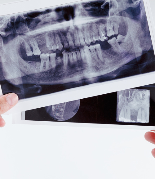
- Panoramic (OPG) x-ray
An OPG is an extraoral x-ray, which means nothing is put in your mouth while it is taken. An OPG gives us a full picture of your mouth and jaw from one side to the other. This is a great screening image as it can check your jaw joint, wisdom teeth, jawbone lesions, unerupted teeth, abnormalities or infection in or around the teeth, and in a general sense the position of your teeth. Having an OPG on record is often recommended, especially for children to assess presence of adult teeth and screen for potential issues, however subsequent OPG would not be taken without a specific need.
- Periapical (PA) x-ray
A periapical x-ray is an intraoral x-ray of usually a specific tooth or area. A small film is placed behind the tooth/area in question. These are similar to a bitewing, however rather than getting half of the top and bottom teeth in the picture, you get all of the top or bottom teeth. For teeth that might need or are in the process of root canals, these are very important to ensure excellent quality treatment. These are never done routinely, only where there is a specific need.
- Cone Beam (CBVT) x-ray
A cone beam is a dimensionally accurate 3-dimensional image. Most commonly, these are used where there is a need to assess areas of the mouth in 3 dimensions, or where accuracy of measurements is important (not possible on 2-d x-rays). Most commonly these are to assess sites for dental implants, (including volume of available bone and proximity to anatomical features such as nerves and adjacent teeth), wisdom teeth, (assessing root anatomy and nerve proximity), jaw lesions (such as a cyst or infection), and even root length and nerve structure within a single tooth. Cone beam x-rays, like periapical x-rays,are never done routinely, and only where there is a specific need.
- Lateral Cephalometric (LatCeph) x-ray
A LatCeph is a lateral view of a person’s whole head. These are used primarily by orthodontists to assess a patient’s jaw positions as they relate to each and to the person’s skeletal base. This will help to design an appropriate and comprehensive orthodontic plan. If you or your child is going to an orthodontist for a consult, they will usually require a LatCeph as well as an OPG.
if you had any further questions, please don’t hesitate to give us a call on (07) 3379 1328